Buscar
Mostrando ítems 111-120 de 176
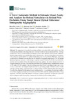
Automatic Identification of Diabetic Macular Edema Using a Transfer Learning-Based Approach
(MDPI AG, 2019-07-31)
[Abstract] This paper presents a complete system for the automatic identification of pathological Diabetic Macular Edema (DME) cases using Optical Coherence Tomography (OCT) images as source of information. To do so, the ...
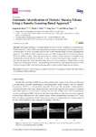
A Novel Automatic Method to Estimate Visual Acuity and Analyze the Retinal Vasculature in Retinal Vein Occlusion Using Swept Source Optical Coherence Tomography Angiography
(MDPI AG, 2019-09-20)
[Abstract] The assessment of vascular biomarkers and their correlation with visual acuity is one of the most important issues in the diagnosis and follow-up of retinal vein occlusions (RVOs). The high workloads of clinical ...
Automatic Wide Field Registration and Mosaicking of OCTA Images Using Vascularity Information
(Elsevier BV, 2019)
[Abstract] Optical Coherence Tomography Angiography (OCTA) constitutes a novel ophthalmological image modality that is characterized for being a non-invasive capture technique that allows a profound analysis of the vascular ...
Multimodal Image Encoding Pre-training for Diabetic Retinopathy Grading
(Elsevier, 2022)
[Abstract] Diabetic retinopathy is an increasingly prevalent eye disorder that can lead to severe vision impairment. The severity grading of the disease using retinal images is key to provide an adequate treatment. However, ...
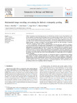
End-To-End Multi-Task Learning Approaches for the Joint Epiretinal Membrane Segmentation and Screening in OCT Images
(Elsevier, 2022)
[Abstract] Background and objectives The Epiretinal Membrane (ERM) is an ocular disease that can cause visual distortions and irreversible vision loss. Patient sight preservation relies on an early diagnosis and on determining ...
Automatic Visual Acuity Estimation by Means of Computational Vascularity Biomarkers Using Oct Angiographies
(M D P I AG, 2019-10-31)
[Abstract] Optical Coherence Tomography Angiography (OCTA) constitutes a new non-invasive ophthalmic image modality that allows the precise visualization of the micro-retinal vascularity that is commonly used to analyze ...
Multi-Stage Transfer Learning for Lung Segmentation Using Portable X-Ray Devices for Patients With COVID-19
(Elsevier BV, 2021-07)
[Abstract] One of the main challenges in times of sanitary emergency is to quickly develop computer aided diagnosis systems with a limited number of available samples due to the novelty, complexity of the case and the ...
Automatic Segmentation and Intuitive Visualisation of the Epiretinal Membrane in 3D OCT Images Using Deep Convolutional Approaches
(IEEE, 2021)
[Abstract] Epiretinal Membrane (ERM) is a disease caused by a thin layer of scar tissue that is formed on the surface of the retina. When this membrane appears over the macula, it can cause distorted or blurred vision. ...
Analysis of Separability of COVID-19 and Pneumonia in Chest X-ray Images by Means of Convolutional Neural Networks
(MDPI AG, 2020-08-21)
[Abstract]
The new coronavirus (COVID-19) is a disease that is caused by severe acute respiratory syndrome coronavirus 2 (SARS-CoV-2). On 11 March 2020, the coronavirus outbreak has been labelled a global pandemic by the ...
Deep Convolutional Approaches for the Analysis of COVID-19 Using Chest X-Ray Images From Portable Devices
(Institute of Electrical and Electronics Engineers, 2020-10-26)
[Abstract]
The recent human coronavirus disease (COVID-19) is a respiratory infection caused by severe acute respiratory syndrome coronavirus 2 (SARS-CoV-2). Given the effects of COVID-19 in pulmonary tissues, chest ...


