Identification of a distinct lipidomic profile in the osteoarthritic synovial membrane by mass spectrometry imaging
Ver/
Use este enlace para citar
http://hdl.handle.net/2183/28505
A non ser que se indique outra cousa, a licenza do ítem descríbese como Creative Commons Attribution-NonCommercial-NoDerivs 4.0 International License (CC-BY-NC-ND 4.0)
Coleccións
- Investigación (FFISIO) [481]
Metadatos
Mostrar o rexistro completo do ítemTítulo
Identification of a distinct lipidomic profile in the osteoarthritic synovial membrane by mass spectrometry imagingData
2021-05Cita bibliográfica
Rocha B, Cillero-Pastor B, Ruiz-Romero C, Paine MRL, Cañete JD, Heeren RMA, Blanco FJ. Identification of a distinct lipidomic profile in the osteoarthritic synovial membrane by mass spectrometry imaging. Osteoarthritis Cartilage. 2021 May;29(5):750-761.
Resumo
[Abstract] Objective: Synovial inflammation is one of the most characteristic events in different types of arthritis, including Osteoarthritis (OA). Emerging evidence also suggests the involvement of lipids in the regulation of inflammatory processes. The aim of this study was to elucidate the heterogeneity and spatial distribution of lipids in the OA synovial membrane and explore their putative involvement in inflammation.
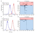
Method: The abundance and distribution of lipids were examined in human synovial membranes. To this end, histological cuts from this tissue were analysed by matrix-assisted laser desorption ionization - mass spectrometry imaging (MALDI-MSI). The lipidomic profile of OA synovium was characterized and compared with healthy and other forms of inflammatory arthropathies as Rheumatoid Arthritis (RA) and Psoriatic Arthritis (PsA) using principal component analysis and discriminant analysis methods. Lipid identification was undertaken by tandem MS analyses and database queries.
Results: Our results reveal differential and characteristic lipidomic profiles between OA and control samples. Specifically, we unveiled that OA synovium presents elevated levels of phosphatidylcholines, fatty acids and lysophosphatidic acids and lower levels of lysophosphatidylcholines compared to control tissues. The spatial distribution of particular glycerophospholipids was also correlated with hypertrophic, inflamed or vascularized synovial areas. Compared with other inflammatory arthritis, the OA tissue showed lower amounts of phosphatidylethanolamine-based plasmalogens.
Conclusions: This study provides a novel insight into the lipid profiles of synovial membrane and differences in abundance between OA and control tissues. The lipidomic alterations improves understanding of the pathogenic mechanisms of OA and may be important for its diagnosis.
Palabras chave
Glycerophospholipids
Mass spectrometry imaging
Osteoarthritis
Synovial membrane
Mass spectrometry imaging
Osteoarthritis
Synovial membrane
Versión do editor
Dereitos
Creative Commons Attribution-NonCommercial-NoDerivs 4.0 International License (CC-BY-NC-ND 4.0)
ISSN
1063-4584


