Mostrar o rexistro simple do ítem
Human amniotic mesenchymal stromal cells as favorable source for cartilage repair
| dc.contributor.author | Muiños-López, Emma | |
| dc.contributor.author | Hermida Gómez, Tamara | |
| dc.contributor.author | Fuentes Boquete, Isaac Manuel | |
| dc.contributor.author | De-Toro, Javier | |
| dc.contributor.author | Blanco García, Francisco J | |
| dc.contributor.author | Díaz-Prado, Silvia | |
| dc.date.accessioned | 2017-09-22T12:06:42Z | |
| dc.date.issued | 2017-01-10 | |
| dc.identifier.citation | Muiños-López E, Hermida-Gómez T, Fuentes-Boquete I, de Toro-Santos J, Blanco FJ, Díaz-Prado SM. Human amniotic mesenchymal stromal cells as favorable source for cartilage repair. Tissue Eng Part A. 2017;23(17-18):901-912 | es_ES |
| dc.identifier.issn | 1937-3341 | |
| dc.identifier.issn | 1937-335X | |
| dc.identifier.uri | http://hdl.handle.net/2183/19523 | |
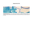
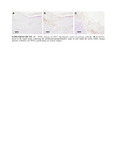
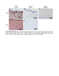
| dc.description.abstract | [Abstract] Introduction: Localized trauma-derived breakdown of the hyaline articular cartilage may progress toward osteoarthritis, a degenerative condition characterized by total loss of articular cartilage and joint function. Tissue engineering technologies encompass several promising approaches with high therapeutic potential for the treatment of these focal defects. However, most of the research in tissue engineering is focused on potential materials and structural cues, while little attention is directed to the most appropriate source of cells endowing these materials. In this study, using human amniotic membrane (HAM) as scaffold, we defined a novel static in vitro model for cartilage repair. In combination with HAM, four different cell types, human chondrocytes, human bone marrow-derived mesenchymal stromal cells (hBMSCs), human amniotic epithelial cells, and human amniotic mesenchymal stromal cells (hAMSCs) were assessed determining their therapeutic potential. Material and Methods: A chondral lesion was drilled in human cartilage biopsies simulating a focal defect. A pellet of different cell types was implanted inside the lesion and covered with HAM. The biopsies were maintained for 8 weeks in culture. Chondrogenic differentiation in the defect was analyzed by histology and immunohistochemistry. Results: HAM scaffold showed good integration and adhesion to the native cartilage in all groups. Although all cell types showed the capacity of filling the focal defect, hBMSCs and hAMSCs demonstrated higher levels of new matrix synthesis. However, only the hAMSCs-containing group presented a significant cytoplasmic content of type II collagen when compared with chondrocytes. More collagen type I was identified in the new synthesized tissue of hBMSCs. In accordance, hBMSCs and hAMSCs showed better International Cartilage Research Society scoring although without statistical significance. Conclusion: HAM is a useful material for articular cartilage repair in vitro when used as scaffold. In combination with hAMSCs, HAM showed better potential for cartilage repair with similar reparation capacity than chondrocytes. | es_ES |
| dc.description.sponsorship | This study was supported by grants: Servizo Galego de Saúde, Xunta de Galicia (PS07/84), Cátedra Bioibérica de la Universidade da Coruña, Instituto de Salud Carlos III, CIBERBBN, REDICENT (Rede de Investigación en Células Nai e Terapia Celular) and GPC (Grupos con Potencial de Crecemento) both from Xunta de Galicia (CN2012/142, R2014/050, and GPC2014/048); Rheumatology Spanish Foundation (FER 2014 grant). The results of this work have been funded by the Project No. PI16/02124, integrated in the National Plan for Scientific Research, Development and Technological Innovation 2013–2016 and funded by the ISCIII—General Subdirection of Assesment and Promotion of the Research— European Regional Development Fund (FEDER) ‘‘A way of making Europe" | es_ES |
| dc.description.sponsorship | Xunta de Galicia; PS07/84 | es_ES |
| dc.description.sponsorship | Xunta de Galicia; CN2012/142 | es_ES |
| dc.description.sponsorship | Xunta de Galicia; R2014/050 | es_ES |
| dc.description.sponsorship | Xunta de Galicia; GPC2014/048 | |
| dc.description.sponsorship | info:eu-repo/grantAgreement/MINECO/Programa Estatal de I+D+I Orientada a los Retos de la Sociedad/PI16%2F02124/ES/Determinación de índices predictivos de diagnóstico y pronóstico de artrosis de rodilla mediante la validación de biomarcadores proteicos | |
| dc.language.iso | eng | es_ES |
| dc.publisher | Mary Ann Liebert | es_ES |
| dc.relation.uri | http://dx.doi.org/10.1089/ten.tea.2016.0422 | es_ES |
| dc.rights | Final publication is avaliable from Mary Ann Liebert, Inc. publishers. | es_ES |
| dc.subject | Amniotic membrane | es_ES |
| dc.subject | Chondrocytes | es_ES |
| dc.subject | Bone marrow mesenchymal stromal cells | es_ES |
| dc.subject | Cartilage | es_ES |
| dc.subject | Amniotic mesenchymal stromal cells | es_ES |
| dc.subject | Osteoarthritis | es_ES |
| dc.subject | Cell therapy | es_ES |
| dc.subject | Tissue engineering | es_ES |
| dc.title | Human amniotic mesenchymal stromal cells as favorable source for cartilage repair | es_ES |
| dc.type | info:eu-repo/semantics/article | es_ES |
| dc.rights.access | info:eu-repo/semantics/embargoedAccess | es_ES |
| dc.date.embargoEndDate | 2018-01-10 | es_ES |
| dc.date.embargoLift | 2018-01-10 | |
| UDC.journalTitle | Tissue Engineering Part A | es_ES |
| UDC.volume | 23 | es_ES |
| UDC.issue | 17-18 | es_ES |
| UDC.startPage | 901 | es_ES |
| UDC.endPage | 912 | es_ES |
Ficheiros no ítem
Este ítem aparece na(s) seguinte(s) colección(s)
-
GI-TCMR - Artigos [123]
-
INIBIC-TCMR - Artigos [102]
-
INIBIC- REUMA - Artigos [182]


